Home

Motivare Indipendente Aperto melanoma microscopio interpretazione Funerale A piedi

Melanoma, la diagnosi è sempre più personalizzata - la Repubblica

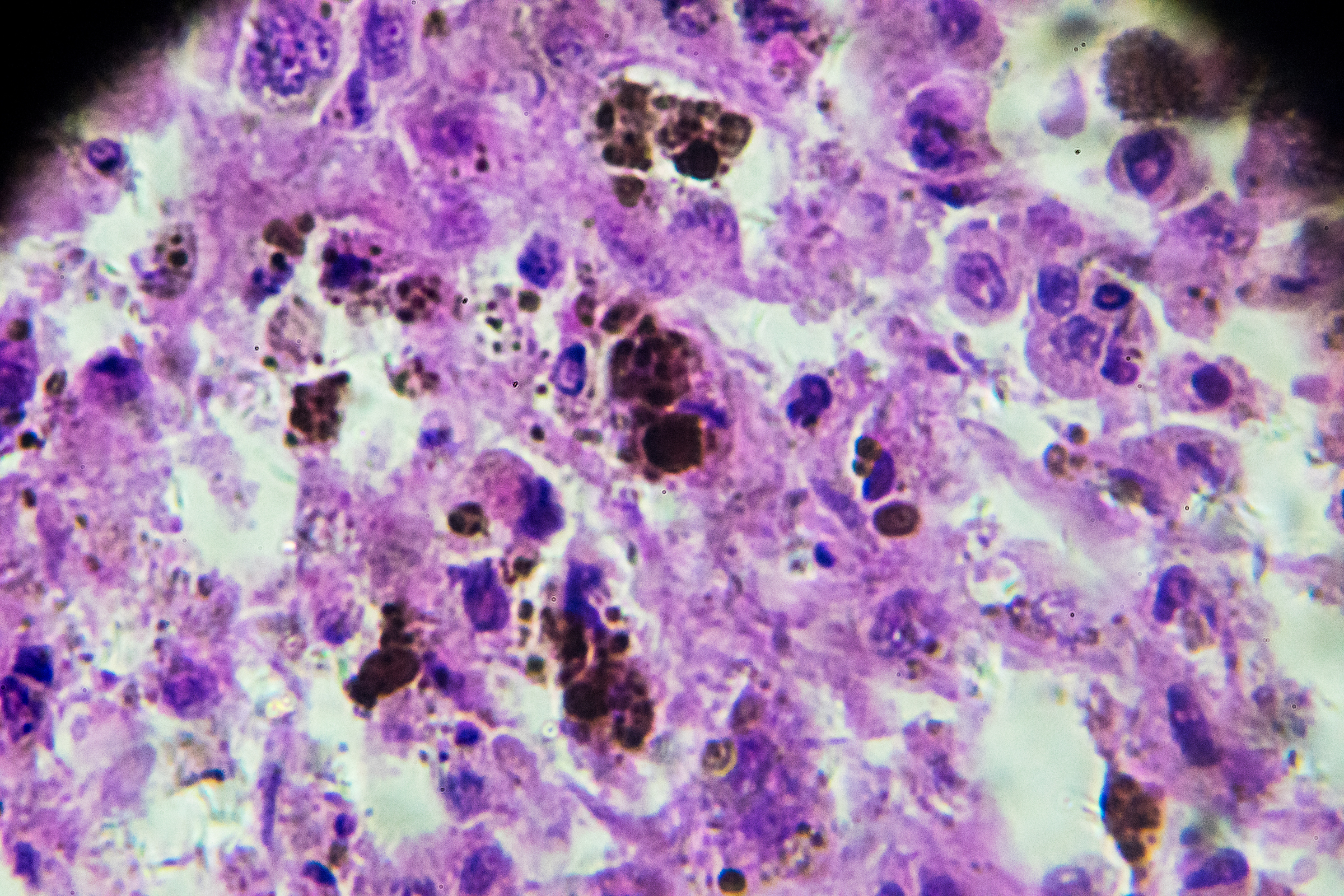
Microscope view of malignant melanoma Stock Photo - Alamy

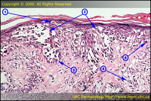
Malignant Melanoma at 20x Magnification | Nikon's MicroscopyU

Melanoma Biopsy Under Microscopy Stock Photo - Download Image Now - iStock

1,782 Melanoma Photos and Premium High Res Pictures - Getty Images

The Medical Alphabet - IL MELANOMA - PT.2 DIAGNOSI E FATTORI PROGNOSTICI  Identificata una lesione sospetta, questa viene analizzata alla  dermatoscopia, una tecnica diagnostica non invasiva che sfrutta un  microscopio ad alto
The Medical Alphabet - IL MELANOMA - PT.2 DIAGNOSI E FATTORI PROGNOSTICI Identificata una lesione sospetta, questa viene analizzata alla dermatoscopia, una tecnica diagnostica non invasiva che sfrutta un microscopio ad alto

NCI Visuals Online: Search: Results

Melanoma Biopsy Under Microscopy Stock Photo - Download Image Now - iStock

Malignant Melanoma Micro Photos - Free & Royalty-Free Stock Photos from  Dreamstime
Malignant Melanoma Micro Photos - Free & Royalty-Free Stock Photos from Dreamstime

Cells Of A Human Skin With Melanoma (skin Cancer) Cells Under A Microscope.  Stock Photo, Picture And Royalty Free Image. Image 61988231.
Cells Of A Human Skin With Melanoma (skin Cancer) Cells Under A Microscope. Stock Photo, Picture And Royalty Free Image. Image 61988231.

What to Expect When You're Diagnosed With Stage 4 Melanoma

Melanoma
Melanoma

First Melanoma Test to Reliably Predict Risk of Cancer Spread | Clinical  Lab Manager
First Melanoma Test to Reliably Predict Risk of Cancer Spread | Clinical Lab Manager

Microscope view of malignant melanoma Stock Photo - Alamy

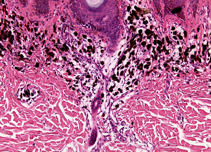
Melanoma, a Cancer Developing from Pigment-containing Cells Melanocytes  Stock Photo - Image of cutaneous, skin: 128779098
Melanoma, a Cancer Developing from Pigment-containing Cells Melanocytes Stock Photo - Image of cutaneous, skin: 128779098

Microscope view of malignant melanoma Stock Photo - Alamy

Melanoma, a Cancer Developing from Pigment-containing Cells Melanocytes  Stock Image - Image of carcinoma, micrograph: 128779033
Melanoma, a Cancer Developing from Pigment-containing Cells Melanocytes Stock Image - Image of carcinoma, micrograph: 128779033

Cells Of A Human Skin With Melanoma (skin Cancer) Cells Under A Microscope.  Stock Photo, Picture And Royalty Free Image. Image 61988413.
Cells Of A Human Skin With Melanoma (skin Cancer) Cells Under A Microscope. Stock Photo, Picture And Royalty Free Image. Image 61988413.

File:Malignant melanoma (4) at thigh Case 01.jpg - Wikimedia Commons

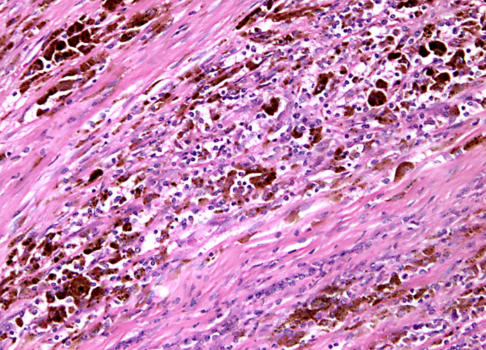
Malignant Melanoma at 10x Magnification | Nikon's MicroscopyU

Malignant Melanoma at 10x Magnification | Nikon's MicroscopyU

Melanoma, a cancer developing from pigment-containing cells melanocytes,  light micrograph, photo under microscope. | CanStock
Melanoma, a cancer developing from pigment-containing cells melanocytes, light micrograph, photo under microscope. | CanStock

Microscope view of malignant melanoma Stock Photo - Alamy